![]()
Imagine being able to view the inner workings of your body, to go beyond the veins, moving into groups of individual cells, exploring their function, their interactions and their evolution over time.
To achieve this, scientists need to first understand how a single cell divides and evolves into a complex organism made up of many cells with differing functions. This is the focus of much current research at the University of Queenland, and Kevin Burrage has just been awarded a five year Federation Fellowship to undertake, in part, two new projects that aim to simulate cellular functions, via mathematical modelling, and to then visualise these functions.
 |
Kevin is working with Perry Bartlett, Rod Rietze (Queensland Brain Institute), Pamela Burrage and Kristin Hatherley to try and understand how precursor or embryonic cells differentiate and evolve into mature functional cells. They are taking single forebrain cells and growing them in vitro into large spherical bodies (neurospheres) consisting of 5-10,000 cells. Kristin (an honours mathematics student) grows neurospheres that express endogenous green fluorescent protein (GFP). Then, using a confocal microscope Kristin develops a series of two dimensional images of the neurosphere interior and using a process called volumetric rendering, gets a three dimensional image of the internal structure of the neurosphere. This allows the researchers to track the migrational history of individual cells. Kristin, Kevin and Pamela are developing mathematical models to understand the process of cell movement and differentiation within the neurosphere. These models will use the cellular automata theory (see Conway’s Game of Life last page) and diffusion equations using partial differential equations (equations involving “differentiation”). The information gained from this research will help Perry and Rod understand cell differentiation and the fundamental process controlling tissue formation during embryogenesis and the continued cellular replacement in tissues of adults. Such information has increasing importance in determining how the adult nervous system retains the ability to produce new functional neurons. |
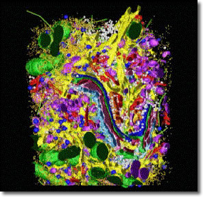
The second project involves researchers in the Centre for Microscopy and Microanalysis, the Institute for Molecular Bioscience (IMB), the Mathematics Department and the Advanced Computational Modelling Centre. The aim here is to develop an immersive three-dimensional model of the interior of a mammalian cell. The idea is to use computational modelling and simulation to link processes occurring inside a cell with images taken from cryo-electron microscopy and high-resolution cell tomography. They will attempt to overlay molecular interaction networks on cellular structures, such as protein trafficking through the Golgi apparatus.
| The aim is to display the visual cell in the University of Queensland’s Virtual Reality Centre http://www.visac.uq.edu.au/ and to allow the user to walk around inside the three dimensional object. Brad Marsh who is to take a position within the IMB from the beginning of 2004 has already made a three dimensional reconstruction of a pancreatic beta cell (see Figure on the right). |
 |